Knee
Dr. Johnson offers the most advanced and innovative treatments in a state-of-the-art facility to maximize patient outcomes.
 Knee
Knee
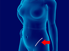
Bikini Incision Hip Replacement
A bikini incision hip replacement is a minimally invasive hip replacement procedure in which the damaged hip joint is replaced with implants through a small diagonal incision made in the front of the hip.

Knee Fracture Surgery
A knee fracture is a broken bone or a crack in or around the joint of the knee. This can involve the tibia (shin bone), the kneecap (patella), or femur (thighbone) where they connect with the knee.

Partial Knee Resurfacing
Partial knee replacement is an alternative to total knee replacement in patients with arthritis on only one side of the knee. Partial knee replacement is a surgical procedure which involves resurfacing and replacement of only the diseased surface of the joint instead of the entire joint.

Quadriceps Tendon Repair
Quadriceps tendon is a thick tissue located at the top of the kneecap. The quadriceps tendon works together with the quadriceps muscles to allow us to straighten our leg. The quadriceps muscles are the muscles located in front of the thigh.

Unicondylar knee Replacement
Unicompartmental knee replacement or unicondylar knee replacement is a minimally invasive surgery in which only the damaged compartment of the knee is replaced with an implant. It is also called a partial knee replacement.

Periprosthetic Knee Fracture Fixation
Periprosthetic knee fracture fixation is a procedure performed to stabilize a fracture that occurs in the bone present around a knee prosthesis. The fracture may involve the lower part of the thighbone (femur), the kneecap (patella) or the upper part of the shinbone (tibia).

ORIF of the Knee Fracture
A knee fracture is a break in the continuity of bone within the knee. This can involve the tibia (shinbone), kneecap (patella), or femur (thigh bone).

Complex Total Knee Replacement
Total knee replacement, also called total knee arthroplasty, is a surgical treatment for painful arthritis of the knee in which the worn-out or damaged surfaces of the knee joint are removed and replaced with an artificial prosthesis.

Rapid Recovery Knee Replacement
Rapid recovery knee replacement, also known as an outpatient knee replacement, is an innovative procedure that is performed to replace a damaged knee joint with a prosthesis using minimally invasive techniques and surgical instruments that minimize post-operative pain and discomfort and promote faster recovery for patients.

Complex Primary Knee Replacement
Complex primary knee replacement is a joint replacement surgery performed on a severely damaged or deformed natural knee joint.

Patellar Tendon Repair
Patellar tendon repair is the surgery performed to reattach the torn tendon to the kneecap and to restore normal function in the affected leg.

Revision Knee Replacement
Revision knee replacement surgery involves replacing a part or all your previous knee prosthesis with a new prosthesis. Although total knee replacement surgery is successful, sometimes the procedure can fail due to various reasons and may require a second revision surgery.

Bilateral Total Knee Replacement
Bilateral knee replacement, also called bilateral knee arthroplasty, is a surgical procedure in which the worn out or damaged surfaces of both knees are removed and replaced with artificial prostheses. Artificial knee joints are usually made of metal, ceramic, or plastic and consist of the femoral and the tibial components.

Robotic Assisted Partial Knee Surgery
Robotic-assisted partial knee surgery is an innovative alternative to the conventional surgical procedure to treat degenerative knee diseases such as osteoarthritis. It is performed using robotic-arm technology that allows your surgeon to precisely perform the surgery through small incisions.

Knee Trauma Reconstruction
Knee trauma reconstruction is a surgical procedure to repair a soft-tissue injury of the knee such as a torn ligament using a tissue graft or replacing the damaged bony surfaces of the knee joint with an artificial knee joint called a prosthesis. Artificial knee joints are usually made of metal, ceramic or plastic, and consist of the femoral and the tibial components.

Correction of a Loose Knee Replacement
Reoperation of a total knee replacement to correct a loosened prosthesis as a result of wear and tear of the prosthetic joint surfaces is known as correction of a loose knee replacement. This procedure involves a complete or partial exchange of prostheses implanted during the original total knee replacement with new prostheses.

Correction of a Painful Knee Replacement
Reoperation of a total knee replacement to resolve a painful knee condition and loss of motion arising out of a damaged or worn out prosthesis is known as correction of a painful knee replacement. This procedure involves a complete or partial exchange of prostheses implanted during the original total knee replacement with new prostheses.

Computer Navigation for Total Knee Replacement
Computer navigation provides your surgeon with real-time 3-D images of your mapped knee and the surgical instruments during surgery. The data for the images is provided by infrared sensors fixed to the bones of the knee and surgical instruments.

Unicompartmental/Partial Knee Replacement
Unicompartmental knee replacement is a minimally invasive surgery in which only the damaged compartment of the knee is replaced with an implant. It is also called a partial knee replacement.

Outpatient Unicondylar Knee Replacement
A unicondylar knee replacement, also known as unicompartmental or partial knee replacement, is a procedure to replace a portion of the damaged knee joint with a prosthetic implant to relieve pain and improve function of the knee joint.

Robotic Assisted Knee Replacement
Robotic-assisted knee replacement surgery is an alternative to the conventional knee replacement procedure. It is performed using robotic-arm technology that allows your surgeon to precisely perform the surgery through a smaller incision as compared to traditional surgery.

Fractures of the Tibia
The lower leg is made up of two long bones called the tibia and fibula that extend between the knee and ankle. The tibia or shinbone is the larger of the two bones. It bears most of the body’s weight and helps form the ankle joint and knee joint.

Knee Arthritis
The joint surface is covered by a smooth articular surface that allows pain-free movement in the joint. Arthritis is a general term covering numerous conditions where the joint surface or cartilage wears out. This surface can wear out for several reasons; often the definite cause is not known. Arthritis often affects the knee joint.
Quadriceps Tendon Rupture
The quadriceps can rupture after a fall, direct blow to the leg and when you land on your leg awkwardly from a jump. Quadriceps tendon rupture most commonly occurs in middle-aged people who participate in sports that involve jumping and running.
Patellar Tendon Rupture
The patellar tendon works together with the quadriceps muscle and the quadriceps tendon to allow your knee to straighten out. Patella tendon rupture is the rupture of the tendon that connects the patella (kneecap) to the top portion of the tibia (shinbone).

Periprosthetic Knee Fractures
Knee replacement, also called knee arthroplasty, is a surgical procedure in which the worn-out or damaged surfaces of the knee joint are removed and replaced with artificial implants. Any resulting fractures or breaks in the bone around the implant are called periprosthetic knee fractures.

Distal Femur Fracture
The femur or thigh bone is the longest and strongest bone in the body, connecting the hip to the knee. A femur fracture is a break in the femur. The distal femur refers to the lower part of the thigh bone which flares out like an upside-down funnel and its lower end is covered by a smooth, slippery articular cartilage that protects and cushions the bone during movement.

Knee Fracture
A fracture is a condition in which there is a break in the continuity of the bone. In younger individuals, these fractures are caused by high energy injuries, as from a motor vehicle accident. In older people, the most common cause is a weak and fragile bone.

Periprosthetic Knee Infection
A very small percentage of patients who undergo joint replacement may develop an infection around the joint. This infection is called a periprosthetic infection.
Quadriceps Tendon Rupture and Repair
A quadriceps tendon rupture is defined as a tear of the quadriceps tendon as a result of a traumatic incident. The quadriceps tendon is a strong rope-like fibrous tissue located at the top of the patella or kneecap that connects the quadriceps muscles to the kneecap.

Patellofemoral Arthritis
Patellofemoral arthritis is an inflammatory condition characterized by loss of the smooth cartilage between the kneecap (patella) and the underlying femoral (thigh) bone in the knee joint. When the articular cartilage wears out, the underlying bones rub against each other, causing pain, swelling, stiffness, and restricted movement.
Knee Anatomy

The knee is a complex joint made up of different structures - bones, tendons, ligaments, and muscles. They all work together to maintain the knee’s normal function and provide stability to the knee during movement.
Having a well-functioning healthy knee is essential for our mobility and ability to participate in various activities. Understanding the anatomy of the knee enhances your ability to discuss and choose the right treatment procedure for knee problems with your doctor.
Bones of the Knee
The knee is a hinge joint made up of two bones, the thighbone (femur) and shinbone (tibia). There are two round knobs at the end of the femur called femoral condyles that articulate with the flat surface of the tibia called the tibial plateau. The tibial plateau on the inside of the leg is called the medial tibial plateau and on the outside of the leg, the lateral tibial plateau.
The two femoral condyles form a groove on the front (anterior) side of the knee called the patellofemoral groove. A small bone called the patella sits in this groove and forms the kneecap. It acts as a shield and protects the knee joint from direct trauma.
A fourth bone called the fibula is the other bone of the lower leg. This forms a small joint with the tibia. This joint has very little movement and is not considered a part of the main joint of the knee.
Articular Cartilage and Menisci of the Knee
Movement of the bones causes friction between the articulating surfaces. To reduce this friction, all articulating surfaces involved in the movement are covered with a white, shiny, slippery layer called articular cartilage. The articulating surface of the femoral condyles, tibial plateaus and the back of the patella are covered with this cartilage. The cartilage provides a smooth surface that facilitates easy movement.
To further reduce friction between the articulating surfaces of the bones, the knee joint is lined by a synovial membrane that produces a thick clear fluid called synovial fluid. This fluid lubricates and nourishes the cartilage and bones inside the joint capsule.
Within the knee joint, between the femur and tibia, are two C-shaped cartilaginous structures called menisci. Menisci function to provide stability to the knee by spreading the weight of the upper body across the whole surface of the tibial plateau. The menisci help in load-bearing i.e. it prevents the weight from concentrating onto a small area, which could damage the articular cartilage. The menisci also act as a cushion between the femur and tibia by absorbing the shock produced by activities such as walking, running and jumping.
Ligaments of the Knee
Ligaments are tough bands of tissue that connect one bone to another bone. The ligaments of the knee stabilize the knee joint. There are two important groups of ligaments that hold the bones of the knee joint together, collateral and cruciate ligaments.
Collateral ligaments are present on either side of the knee. They prevent the knee from moving too far during side to side motion. The collateral ligament on the inside is called the medial collateral ligament (MCL) and the collateral ligament on the outside is called the lateral collateral ligament (LCL).
Cruciate ligaments, present inside the knee joint, control the back-and-forth motion of the knee. The cruciate ligament in the front of the knee is called anterior cruciate ligament (ACL) and the cruciate ligament in the back of the knee is called posterior cruciate ligament (PCL).
Muscles of the Knee
There are two major muscles in the knee - the quadriceps and the hamstrings, which enable movement of the knee joint. The quadriceps muscles are located in front of the thigh. When the quadriceps muscles contract, the knee straightens. The hamstrings are located at the back of the thigh. When the hamstring muscles contract, the knee bends.
Tendons of the Knee
A tendon is a tissue that attaches a muscle to a bone. The quadriceps muscles of the knee meet just above the patella and attach to it through a tendon called the quadriceps tendon. The patella further attaches to the tibia through a tendon called the patella tendon. The quadriceps muscle, quadriceps tendon, and patellar tendon all work together to straighten the knee. Similarly, the hamstring muscles at the back of the leg are attached to the knee joint with the hamstring tendon.
If you wish to be advised on the most appropriate treatment, please call to schedule an appointment or click here to request an appointment online.